
Monthly newsletter
Issue 004 - June 2026
We have had some very good cases at SCEH this month, particularly with some interesting and technically challenging surgical cases – from orthopaedic disease to gastric impactions to abdominal hernias fixed with mesh.
Medial malleolus fracture in trail riding horse
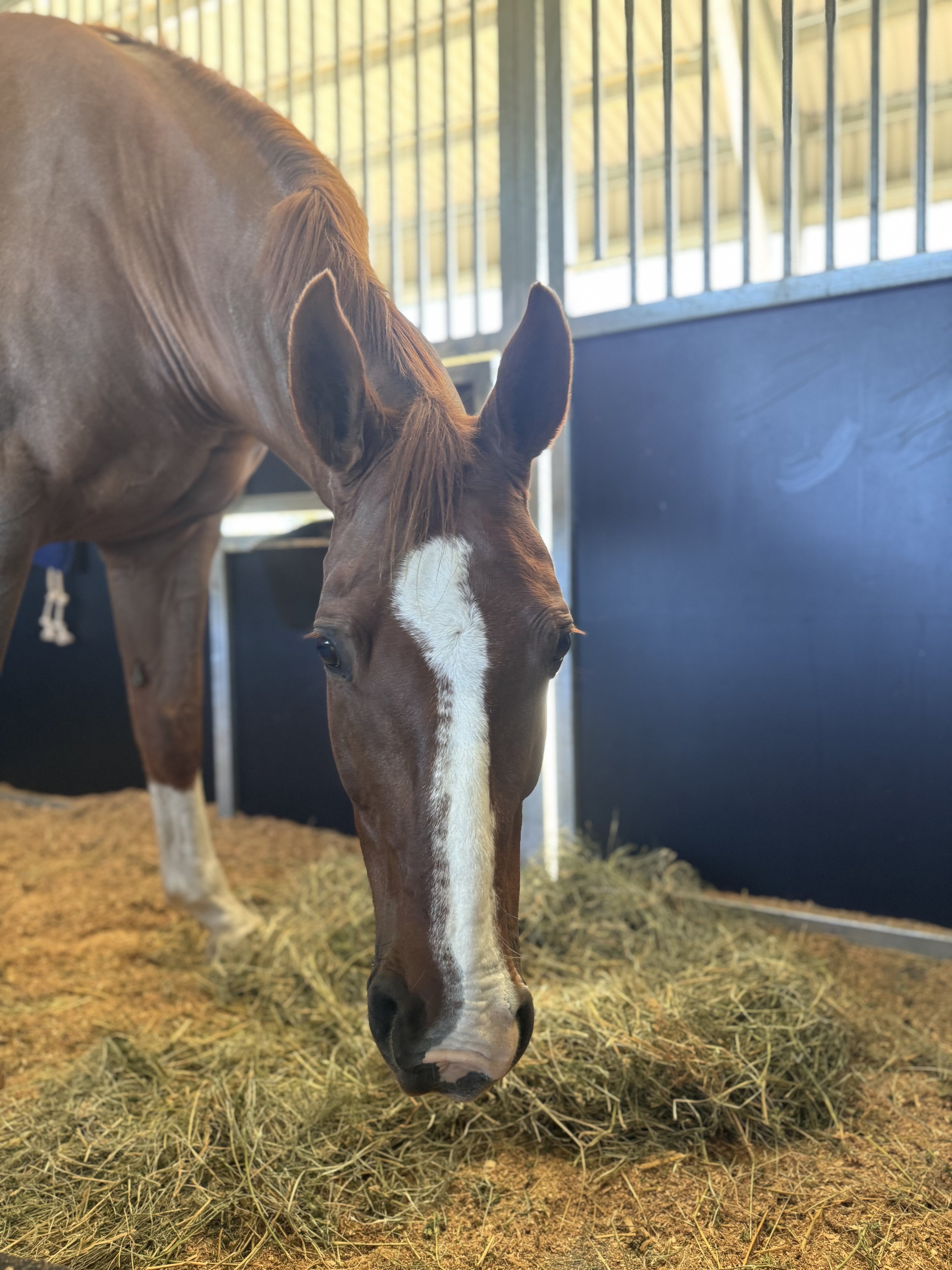
This month, Dr Bridget Nottle and the team treated Chevy, a 9-year-old Quarter Horse gelding, for a chronic fracture involving the medial malleolus of his left hock. Chevy is a trail riding horse along Noosa North Shore.
The medial malleolus is a bony prominence located on the inside of the hock, and fractures in this region can lead to ongoing joint inflammation, discomfort, and reduced performance if left untreated. It is unusual to have fractures off the medial malleolus that have not been associated with a traumatic incident, and that are also articular.
Radiographs identified a large chronic fracture fragment that had separated from the medial malleolus and was contributing to ongoing inflammation and changes within the joint. Following assessment, the decision was made to proceed with surgery to remove the fragment and address the associated pathology.
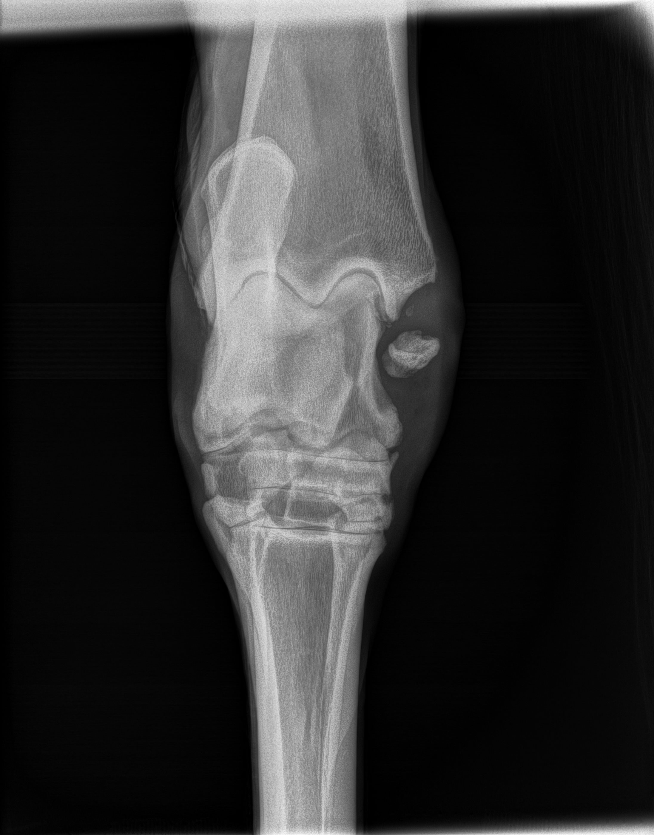
Pre-operative radiograph showing the fracture fragment from the medial malleolus
Chevy underwent arthroscopic surgery under general anaesthesia. Arthroscopy allows surgeons to examine and treat pathology within a joint using a camera and specialised instruments inserted through small incisions.
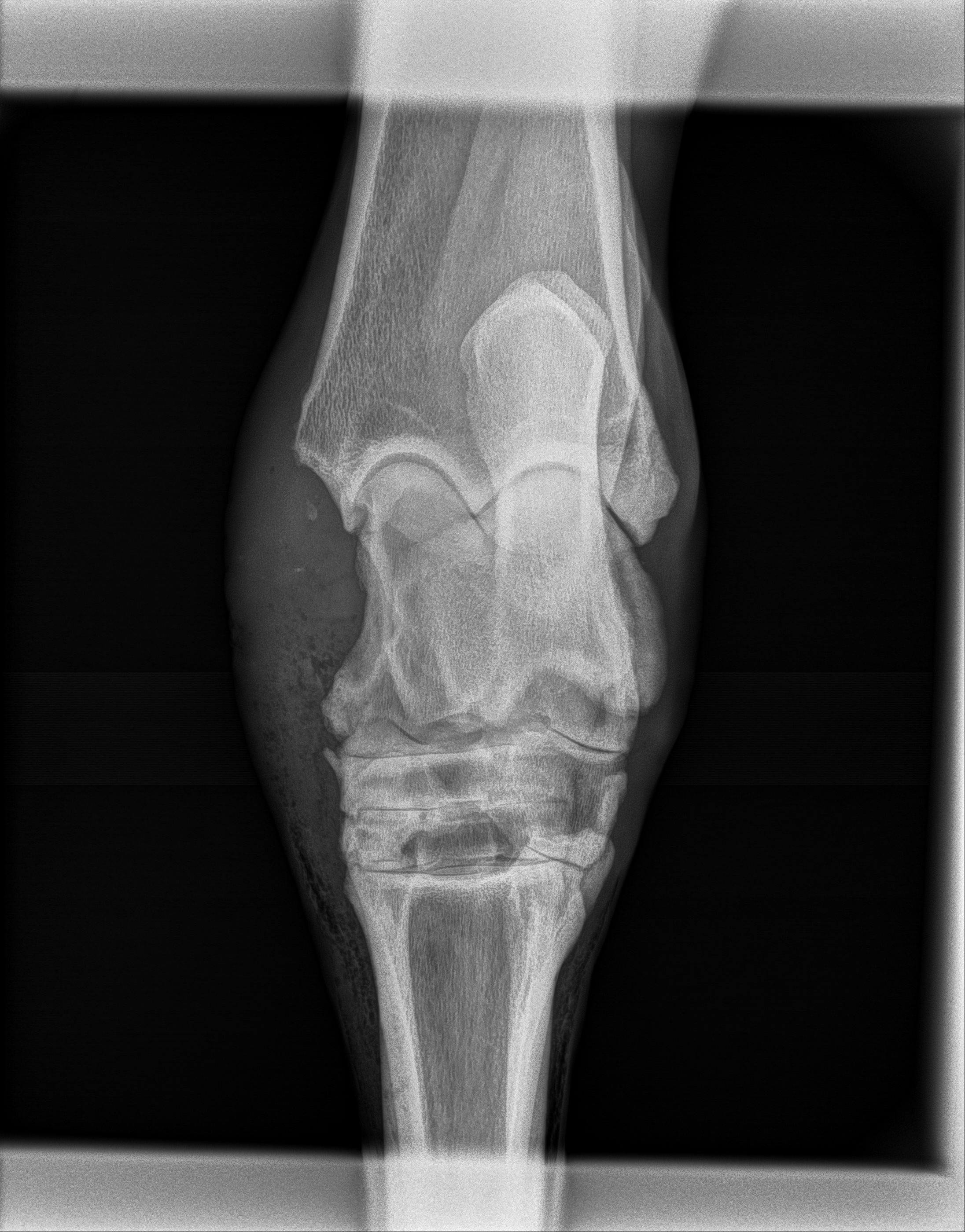
Intra-operative radiograph showing removed fracture fragment
Once inside the joint, significant synovial proliferation and fibrosis were identified. These changes occur when chronic inflammation stimulates thickening of the joint lining and development of excessive fibrous tissue. The abnormal tissue was carefully debrided to improve the joint environment.
Due to the size and location of the fracture fragment, an additional surgical approach was required to safely access and remove it. The fragment was successfully extracted, and the joint was thoroughly assessed before closure.
Chevy recovered smoothly from anaesthesia and remained in hospital for several days while the team monitored his incisions, bandages, comfort, and recovery. He has now returned home to begin a period of stall rest and controlled exercise before gradually progressing through his rehabilitation program over the coming months.
Cases such as Chevy's highlight the value of investigating persistent lameness and performance concerns early. Chronic fracture fragments within joints can result in ongoing inflammation and progressive degenerative joint disease, but advanced imaging and surgical intervention can often provide horses with a much more comfortable future.
We look forward to following Chevy's recovery and wish him all the best on his journey back to leading rides along the picturesque Noosa North Shore.
Gastric impaction causing colic
This month we had a 3-year-old Warmblood gelding presented to Dr Brianna for acute onset severe colic. After thorough diagnostic workup it was identified the gelding had a large gastric impaction.
Gastric impactions occur when feed material persists in the stomach for a prolonged period of time. They can occur in isolation or in other gastrointestinal diseases (such as colon disorders). There is actually not a very good understanding of why gastric impactions occur with 3 broad causes – obstruction with foreign material, mechanical issue with the emptying into the small intestine and function issue with contraction of the stomach to empty food.
In this gelding’s case his stomach was lavaged over 48 hours, he was given intravenous fluids and drugs to help motility of the stomach. We were able to empty the stomach and reintroduced him to low-bulk feed slowly. Further investigations are ongoing with this gelding to identify for the cause of his stomach abnormality and have included gastroscopies and biopsies.
Gastric impactions can be very frustrating for both owners and veterinarians and show that every case is different and will need tailored treatments and diagnostics.

Gastroscope image showing the gastric impaction
Fixing hernias with mesh
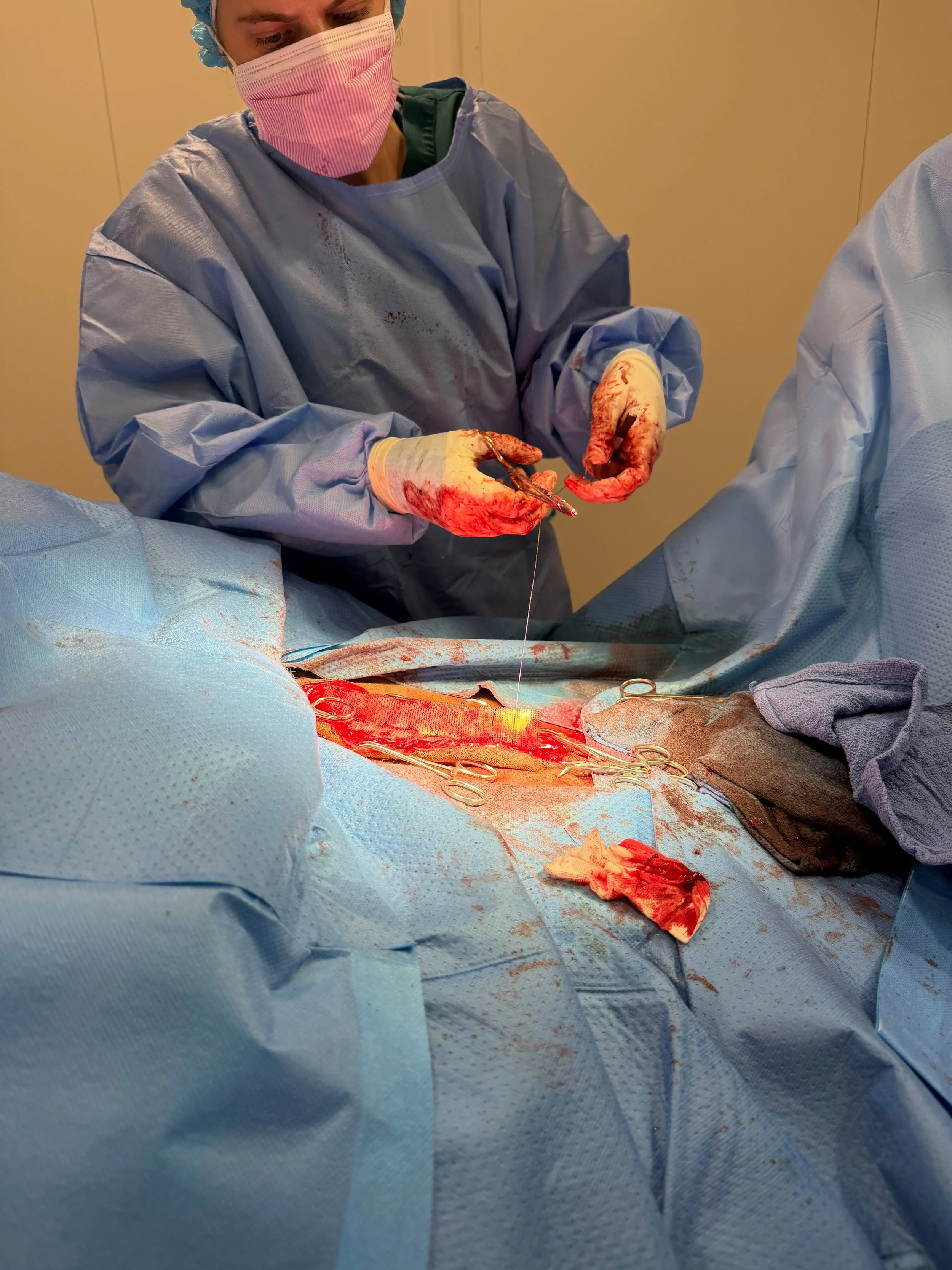
This month we had a 608kg gentle giant, 10-year-old Warmblood referred in for repair of a large abdominal hernia of the linea alba. The owner’s wanted to get him back eventing, thus surgical repair of the hernia was advised.
Given the large hernia and the difficulty in pulling the sides together, Dr Bridget opted for a prosthetic mesh to repair the abdominal defect with an onlay technique. The surgery itself is very time consuming, so consideration to anaesthesia time is very critical, especially with a large horse. Also, another major consideration is the short and long-term acceptance of the body for the prosthesis and other complications can occur such as infection.

We are happy to report that the gelding is so far recovered well from surgery and will go back to work in a few months time. A great result for the horse and owner!
We thank all our clients for their ongoing support and entrusting us with the care of their horses.
The SCEH team.