
Newsletter - Sept ‘26
Issue 007 - September 2026
We’ve had another busy month at Sunshine Coast Equine Hospital, with some particularly interesting medical and surgical cases coming through our doors.
This month we’re sharing three very different cases: a horse with acute kidney injury following an adverse reaction to medication, a tiny foal requiring major abdominal surgery to bypass a narrowed stomach outlet, and a mare with a significant ligament injury who is making great progress with rehabilitation.
Read on for a look at some of the cases we’ve been managing at SCEH this month.
Acute kidney injury in a dressage horse
An11-year-old Warmblood gelding competing at high level FEI dressage was referred to SCEH on a Sunday afternoon after becoming lethargic and showing mild signs of colic. In the days before referral, he had been treated for a skin condition with antibiotics and non-steroidal anti-inflammatory drugs (NSAIDs). On arrival, Drs Tias and Mikarla found him dehydrated, with very high kidney values on bloodwork and a mild impaction of the pelvic flexure of the large colon. He was started on intravenous fluids and monitored closely.
The following day, Dr Brianna performed repeat assessment, including ultrasound examination. Both kidneys were mildly enlarged, supporting a diagnosis of acute kidney injury. The enlarged kidneys were likely contributing to his abdominal discomfort, while the dehydration had probably contributed to the mild colonic impaction.
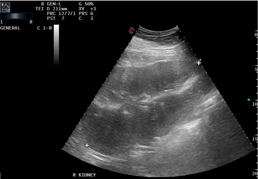
Transabdominal ultrasound of the right kidney
Acute kidney injury isn’t common in horses, but when it occurs it can be serious. There are several potential causes, including severe systemic illness, kidney infections and immune-mediated disease. One important cause is exposure to drugs that can be harmful to the kidneys, particularly when a horse is already unwell or dehydrated. These include medications such as gentamicin, flunixin meglumine, phenylbutazone, oxytetracycline, doxycycline and meloxicam. Importantly, these are commonly used medications and are generally safe when used appropriately. The risk to the kidneys increases when a horse is dehydrated or otherwise unwell, which is why careful monitoring and veterinary guidance are so important.
For this gelding, treatment focused on giving the kidneys the best possible environment to recover. He received intravenous fluids, while potentially kidney-damaging medications were avoided. Bloodwork was repeated regularly to monitor his kidney values, along with frequent urine testing to assess kidney function. The first few days were touch and go. He was dull, had a poor appetite and was certainly not feeling himself. As his kidney values improved, however, so did his appetite and attitude. After a week in hospital, he was well enough to go home.
A repeat ultrasound showed that his kidneys had returned to normal size, and at subsequent rechecks his kidney function continued to improve. He is now back to his usual boisterous self, although he continues to rest from exercise to allow his kidneys time to fully recover.
There is still a possibility that an episode of acute kidney injury can leave some degree of lasting damage, so we will continue to monitor him. For now, though, he has made a very encouraging recovery.
A tiny foal and major abdominal surgery
One quite notable case through SCEH this month was a two-week-old foal who required major abdominal surgery. He had initially been hospitalised as a four-day-old foal with a meconium impaction and had gone home doing well. A short time later, he returned to SCEH with sudden excessive drooling, teeth grinding and reduced nursing.
As part of his investigation, Dr Brianna performed a gastroscopy. This revealed ulceration of both the oesophagus and the squamous lining of the stomach (images below), but the most significant finding was at the pylorus – the opening between the stomach and small intestine. It was extremely narrowed, allowing very little milk to pass through (picture below with arrow). The foal had developed pyloric stenosis, with secondary reflux oesophagitis. Essentially, his stomach could no longer empty normally, so the contents were backing up into the oesophagus and causing significant irritation and ulceration.
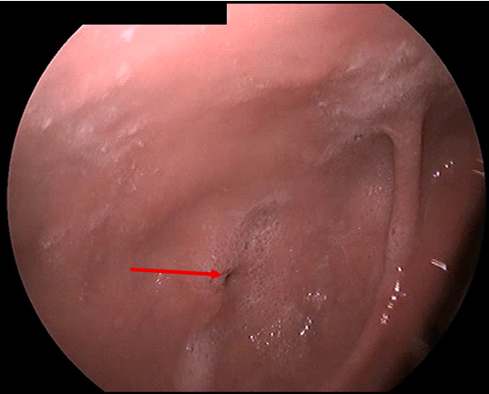
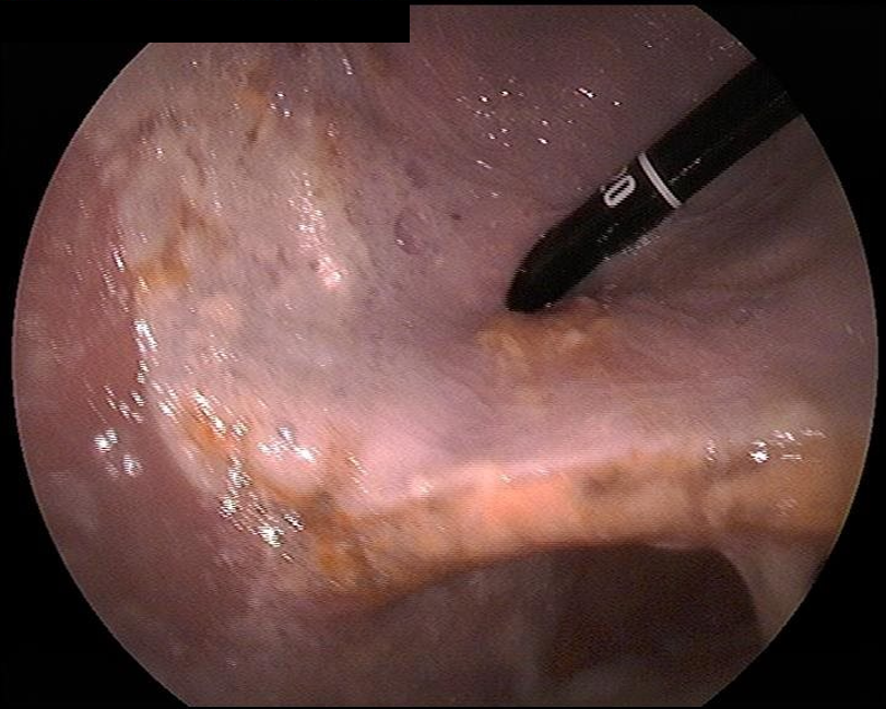
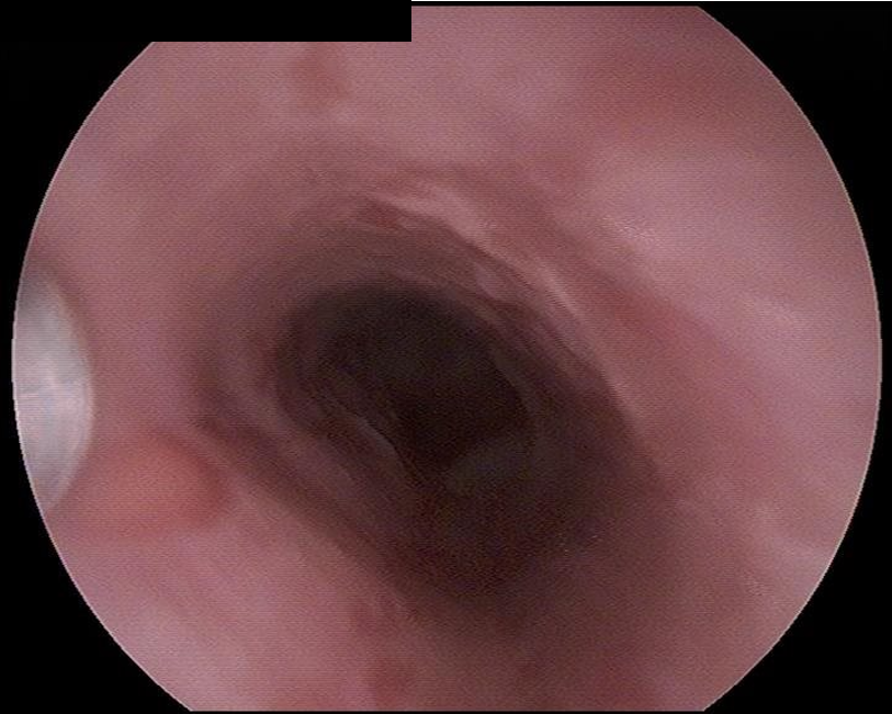
Pyloric stenosis can occur as part of a condition known as gastroduodenal ulcer syndrome in foals. Damage around the pylorus can lead to scar tissue and fibrosis, progressively narrowing the opening. Foals may show signs of colic, reflux, poor growth or failure to thrive because they cannot empty their stomach or absorb nutrients normally. The exact initial cause is not always known, although the condition appears to be more common in foals that have been unwell earlier in life.
Once the narrowing becomes severe, surgery is generally the only option. Rather than trying to open the narrowed pylorus, the surgeon creates a new route for the stomach contents to pass into the small intestine. In other words, the stomach is essentially “rerouted” around the problem area. This is a major procedure in a very small patient, and the risks do not end when the surgery is finished. Careful anaesthesia, intensive post-operative monitoring and very gradual reintroduction of milk are all critical.
Dr Bridget performed the surgery, with Dr Mikarla managing the anaesthesia. The procedure was successful and the foal was transferred straight back to our ICU, where Dr Brianna and the nursing team provided round-the-clock care. He received intravenous fluids and medications to support gastrointestinal motility, and feeding was reintroduced very cautiously. For the first 36 hours, he was carefully refed, with close monitoring for any signs of reflux or digestive complications. Thankfully, he tolerated his feeds extremely well and continued to improve. Ten days after surgery, he was able to head home with his mum.
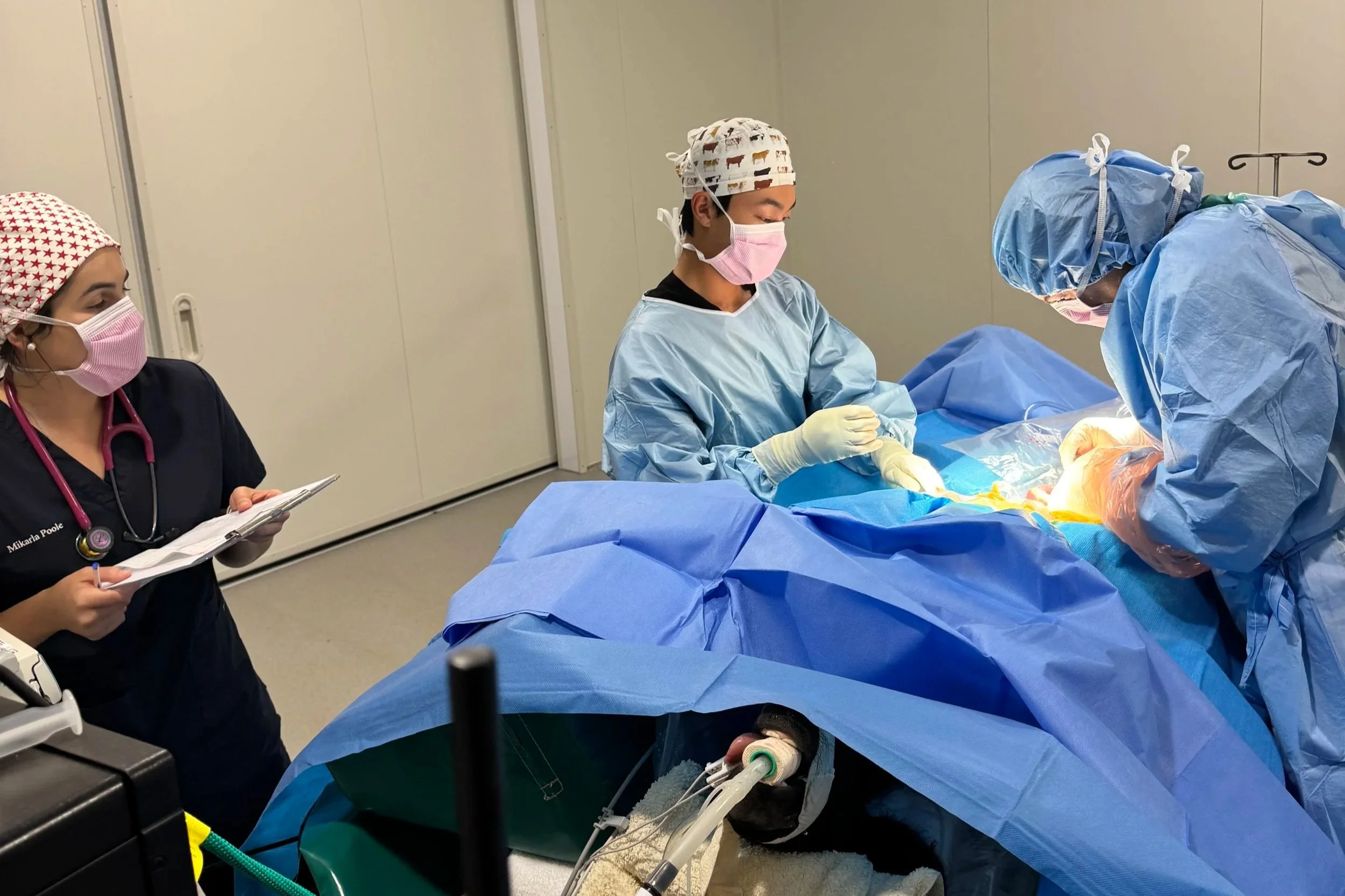
The anaesthetic (Dr Mikarla) and surgical (Dr Bridget and assistant Jun) performing major abdominal surgery on the foal
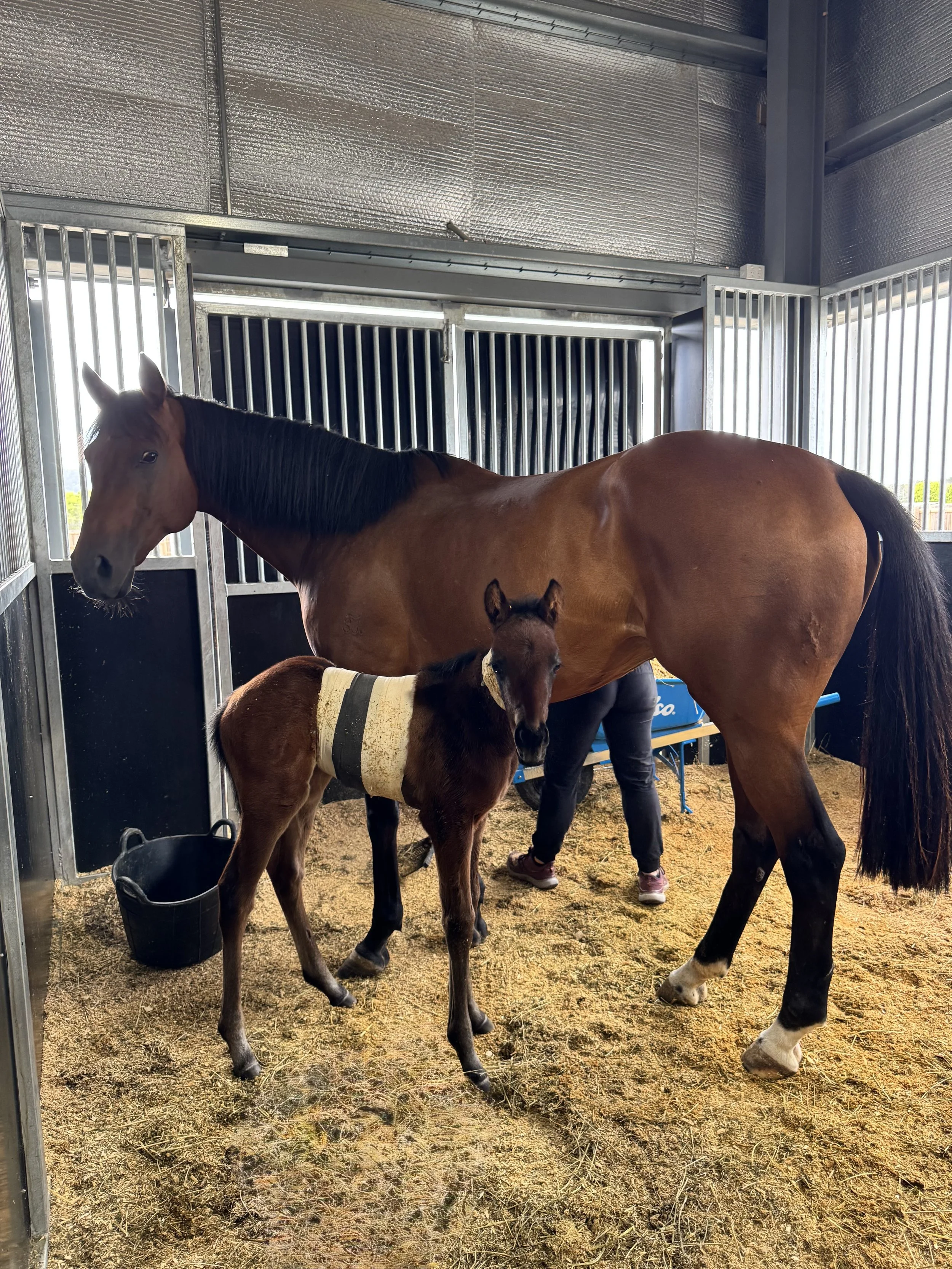
The foal the day of discharge
He has continued to do well, which is a fantastic result for this little colt. Cases like this really highlight the importance of having an experienced surgical, medical and nursing team working together, particularly when caring for critically ill neonatal foals.
Multimodel management for ligament injury
A 16-year-old Clydesdale cross mare was recently presented to Dr Bridget for ongoing right forelimb lameness. There was marked swelling of the lower limb and she was moderately lame at the trot. Ultrasound examination revealed a significant injury to the inferior check ligament, with approximately 60–80% of the ligament fibres torn.
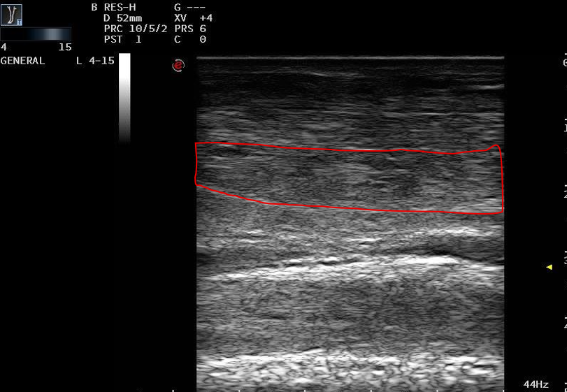
Ultrasound image showing disruption of normal fibre pattern of the inferior check ligament.
The inferior check ligament is an important supporting structure associated with the deep digital flexor tendon. Like tendon injuries, ligament injuries heal through a series of stages, from initial inflammation through to tissue repair and, finally, remodelling. That final stage is particularly important. New fibres need time and controlled exercise to remodel and align correctly, which is why returning a horse to work too quickly can result in reinjury.
For this mare, a strict, structured rehabilitation program was prescribed. Dr Bridget also performed extracorporeal shockwave therapy every two weeks for three treatments, alongside appropriate nutrition and supplementation. Six weeks later, repeat ultrasound showed a remarkable improvement, with the ligament structure approaching normal with clear evidence of healing.
It is still important to remember that a good ultrasound at six weeks does not mean the ligament is ready for full work. Continued rehabilitation and a gradual return to exercise are essential to give the tissue the best chance of developing appropriate strength and alignment.
This case is a great reminder that early investigation of soft tissue injuries is important. With the right diagnosis, patience and a carefully managed rehabilitation program, horses with significant ligament injuries can have a good chance of returning to comfortable work.
Another busy month wrapped up at SCEH! We love sharing some of the interesting cases that come through our doors and giving you a little insight into what goes on behind the scenes at an equine hospital.
Thank you for your continued support as we grow. We look forward to sharing more horse health stories with you next month!